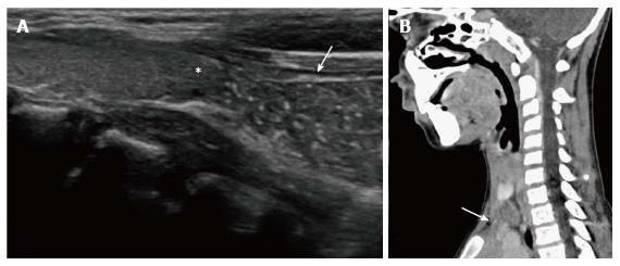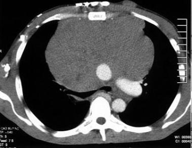

Some CT imaging parameters were significantly correlated with stage, classification and the completeness of resection of thymomas, but none can reliably predict overall survival or disease-free survival duration. Moreover, CT does offer a higher sensitivity and specificity for detecting of thymic lesions regardless of high radioactivity. Ĭomputed tomography (CT) is currently the first choice to identify and characterize thymic masses due to the high spatial, temporal resolution and convenience.

Imaging evaluation has been employed to initially diagnose and properly staging thymoma, especially, on the detection of direct invasion and distant organ metastasis. Therefore, patients would be overtreated or be in lack of essential therapy if thoracic surgeons could not differentiate the properties of thymic lesions. Generally, surgery is often performed under patients with paraneoplastic syndromes like myasthenia gravis or with thymic malignancy except for lymphoma. Thymectomy is the main therapeutic method of thymic masses, while radiotherapy and chemotherapy are often as (neo)adjuvant and palliative procedures. Thymic carcinomas are far rarer but more aggressive than thymomas, thus thymic carcinomas is often found having invaded to adjacent structures and metastasized to distant organ. The overall incidence of thymoma is 1.5 cases per million ( Thymoma is a slow-growing neoplasm that can invade to adjacent structures, including the pericardium and pleura, whereas distant metastases are rare. Although TETs are rare neoplasms as to the whole tumors, they are the most common mediastinal tumors in adults, including thymomas and thymic carcinomas. Thymic cysts are relatively uncommon lesions that can be found at any age and can be congenital or acquired.

True thymic hyperplasia is usually regarded as a rebound phenomenon and characterized by an increase in mass of the gland after a stressor, such as chemotherapy, radiation, steroid treatment, burns or surgery.

Thymic masses are comprised of hyperplasia, cysts, thymic epithelial tumors (TETs), lymphomas, malignant germ cell tumors or metastatic cancers and so on. Keywords: thymic mass, thymomas, CT, MRI, diagnostic accuracy Introduction Ĭonclusion: The diagnostic accuracy of MRI is superior to CT in detecting thymomas, thymic cysts or thymic hyperplasia but that of CT and MRI is still unclear in differentiating thymic carcinomas and lymphomas/germ cell tumors. AUC of CT was 0.875 and that of MRI was 0.880. The sensitivity of CT and MRI was both 100%, while the specificity was 75% and 80%, respectively. We showed outcomes of quantitative analysis of each study in this article. There were 253 cases examined by CT and 340 cases by MRI in total. Results: Eight literatures were finally included and analyzed in this study. The ROC curve was applied to compare the diagnostic performance of different imaging modalities. Methods: We searched literature and collected information on first author, publication year, cases of different types of thymic lesions, correct diagnostic cases of CT and MRI and results of quantitative analysis of CT and MRI. Purpose: The aim of this study was to compare diagnostic accuracy between CT and MRI for thymic masses. Select the file that you have just downloaded and select import option Reference Manager (RIS).
#Residual thymic tissue download
Available fromĬlick on Go to download the file. Comparison between CT and MRI in the Diagnostic Accuracy of Thymic Masses.


 0 kommentar(er)
